Quick Summary
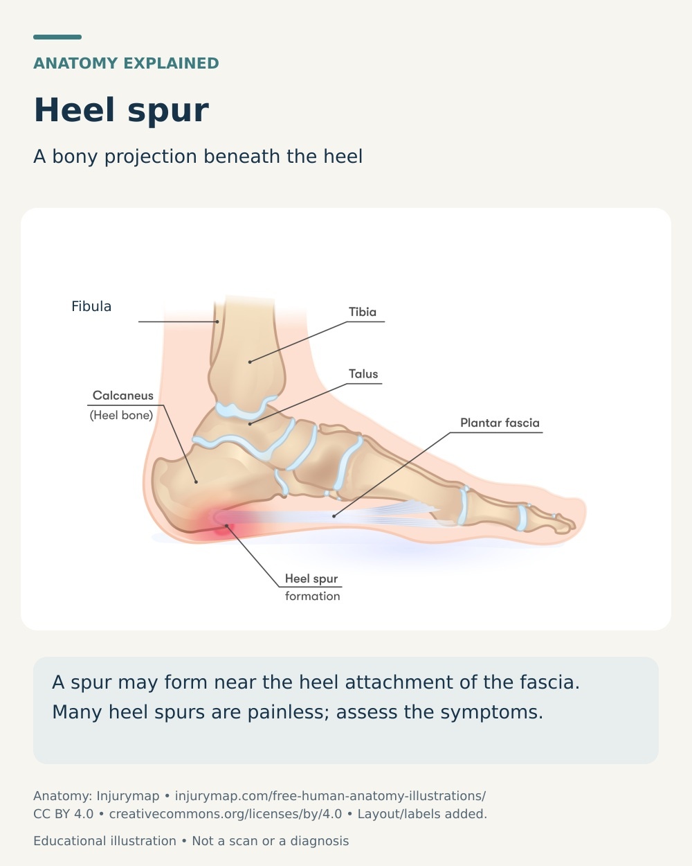
A heel spur is a bony growth on the heel bone (calcaneus), most often on the underside where the plantar fascia attaches. Research shows the spur itself is usually not the source of pain — discomfort typically comes from the surrounding soft tissue and from how your heel is loaded — so our care targets those factors rather than the bone.
At our New Malden clinic we combine hands-on manual therapy, a targeted exercise programme and, for persistent cases, radial shockwave therapy using the Swiss Storz Medical MASTERPULS. Research suggests shockwave may help reduce chronic heel pain, typically over 3–6 sessions.
What Is a Heel Spur?
A heel spur (calcaneal spur) is a bony outgrowth that forms on the calcaneus, the largest bone in your foot. It develops where soft tissue attaches to the bone, and there are two distinct types.
Plantar (underside) heel spur
This forms on the underside of the heel bone, close to where the plantar fascia and the small muscles of the foot attach. It is the type most often linked with pain under the heel and with plantar fasciitis.
Posterior (back-of-heel) spur
This forms at the back of the heel where the Achilles tendon inserts, and is associated with insertional Achilles tendinopathy. It tends to cause pain at the back of the heel, often aggravated by the firm heel counter of a shoe.
Spurs are thought to develop through repeated traction and loading at these attachment points, with gradual degenerative change over time.
(Kirkpatrick et al., Journal of Anatomy, 2017)
A spur on an X-ray is often not the cause of pain
Heel spurs become more common with age — reported in around 11–21% of younger adults, rising to about 55% in those over 62. They are found in 45–85% of people with plantar fasciitis, but also in between 10% and 63% of people who have no heel pain at all. A radiographic study of 1,335 patients found plantar spurs in 32.2% and posterior spurs in 13.1%, both increasing with age.
(Kirkpatrick et al., Journal of Anatomy, 2017; Toumi et al., BMC Musculoskeletal Disorders, 2014)
Because spurs are so common in pain-free heels, your symptoms and examination findings matter far more than what appears on an X-ray. In most cases the pain arises from the plantar fascia or the Achilles insertion rather than from the bone itself.
The outlook is generally encouraging. Most people with plantar heel pain improve with conservative care, although recovery can be gradual, with evidence suggesting symptoms settle in around 60–80% of patients within 12 to 24 months.
Do You Experience These Symptoms?
✓ Sharp or deep pain under the heel, or at the back of the heel
✓ Pain that is worst with your first few steps in the morning or after sitting, easing after a few minutes of walking
✓ Pain that returns after prolonged standing or towards the end of the day
✓ Tenderness when you press firmly on the inner front of the heel, or over the Achilles insertion
✓ Discomfort that increases with walking, running or standing on hard surfaces
✓ A tendency to walk on your forefoot or tiptoes to take pressure off the heel
✓ Aggravation from the rigid heel of certain shoes, particularly with a back-of-heel spur
These symptoms can be treated.
The sooner you begin, the better your chances of a smoother recovery.
What Causes a Heel Spur?
• Repetitive traction and overload
Chronic tension where the plantar fascia or Achilles tendon meets the heel bone is the leading explanation for spur formation.
Over time this repeated loading is associated with degenerative change and bony growth at the attachment.
• Age and accumulated loading
Spurs become considerably more common with age, reflecting years of cumulative mechanical stress through the heel.
This is why they are so often found as incidental discoveries in older adults with no heel pain whatsoever.
• Body weight and foot mechanics
A higher body weight, altered foot posture and excessive pronation all increase the load transmitted through the heel attachments.
These same factors are strongly associated with plantar fascia problems, which frequently occur alongside a spur.
• Activity and footwear
A recent increase in running, jumping or standing on hard surfaces can overload the heel more quickly than the tissue can adapt.
Unsupportive or worn-out footwear adds to this, and for back-of-heel spurs a tight heel counter can be a specific aggravator.
• Inflammatory joint conditions
Conditions such as ankylosing spondylitis, psoriatic arthritis and reactive arthritis can drive spur formation through inflammation at the tendon attachment.
These spurs may affect both heels, and managing the underlying condition alongside physiotherapy is important.
How We Treat Heel Spurs at J&J Therapy
Hands-On Manual Therapy
Our HCPC-registered physiotherapists use hands-on manual therapy — including soft-tissue release, joint mobilisation and specific calf and plantar fascia techniques — to reduce pain and restore movement around your heel and ankle. Because the bony spur itself cannot be reshaped by hand, we target the surrounding soft tissue, calf tightness and ankle mobility that influence how your heel is loaded. Evidence shows hands-on treatment works best as part of a combined programme rather than as a stand-alone therapy, so it is always paired with exercise.
Shockwave Therapy*
Swiss Storz Medical MASTERPULS radial shockwave delivers acoustic pressure waves to the painful heel attachment. Research suggests this may reduce chronic heel pain and stimulate healing in the soft tissue around the spur, particularly when combined with loading exercises. It does not dissolve or remove the bone itself — it targets your symptoms and the surrounding tissue. Non-invasive, evidence-based. Typically 3–6 sessions.
Targeted Exercise Programme
Appropriate loading is central to recovery, so your physiotherapist will prescribe an individualised, progressive exercise programme. Research suggests high-load strength work, such as slow single-leg heel raises with a towel under the toes, may improve pain and function in the early months compared with stretching alone. We combine calf and plantar fascia loading, ankle mobility work and a graded return to walking, running or standing, tailored to whether your spur is on the underside or the back of the heel. For back-of-heel spurs we avoid aggressive stretching that can irritate the Achilles insertion.
When to Seek Urgent Medical Help
While most symptoms are not serious, urgent medical attention is required if you experience any of the following:
Heel pain after a fall or a sudden increase in running or impact activity, especially if you cannot put weight through the foot — this may indicate a stress fracture
Heat, redness, swelling or fever in the heel, or heel pain alongside pain and stiffness in other joints, your lower back or your eyes
Burning pain, pins and needles, numbness or electric-shock sensations in the heel or sole, which may suggest nerve involvement
A sudden pop at the back of the heel with an inability to walk normally or push off, which needs urgent assessment
FAQs About Heel Spurs
Can a heel spur be removed or dissolved without surgery?
Physiotherapy, exercise and shockwave therapy do not dissolve or remove the bony spur. The reassuring news is that this usually does not matter, because the spur itself is rarely the source of pain — the surrounding soft tissue and the way your heel is loaded are.
Treating those factors settles symptoms for most people without any need to address the bone. Surgery to remove a spur is uncommon and reserved for specific, persistent cases.
What is the difference between a heel spur and plantar fasciitis?
Plantar fasciitis is irritation and degeneration of the plantar fascia, the band of tissue running under your foot, and it is the most common cause of heel pain. A heel spur is a bony growth at the point where that fascia attaches to the heel bone.
The two often occur together, but the pain typically comes from the fascia rather than the spur, and many people with a spur have no pain at all.
Do I need an X-ray or a scan to diagnose a heel spur?
Usually not. Heel pain is diagnosed clinically from your history and examination, and imaging is not routinely required.
Because a spur is such a common incidental finding, an X-ray showing one rarely changes how we manage your symptoms. Your physiotherapist may recommend imaging only if your presentation is unusual or to rule out other causes such as a stress fracture.
Does shockwave therapy get rid of the heel spur?
Not exactly, and we think it is important to be clear about that. Research shows shockwave therapy is used to reduce chronic heel pain and to stimulate healing in the soft tissue around the heel, but the best-controlled studies show it does not reliably shrink or remove the bony spur itself.
Because the spur is usually not the true source of pain, reducing your symptoms and improving how your heel is loaded is what matters. We use the Swiss Storz Medical MASTERPULS radial device, typically over 3–6 sessions, alongside your exercise programme. Shockwave is not suitable for everyone, so our physiotherapists will assess you first.
What footwear or insoles help with a heel spur?
Supportive, cushioned shoes with a firm heel counter and good arch support usually help, and gel heel pads or arch-support insoles can take pressure off the painful area.
Try to avoid walking barefoot on hard surfaces and replace worn-out footwear. If your spur is at the back of the heel, choose shoes that do not press on the Achilles insertion. Your physiotherapist can advise on what suits your foot type and activity.
This page is for general information only and does not replace professional medical advice.
Reviewed by Lavya Arigalayan, MSc, HCPC Registered Physiotherapist.
Last reviewed: