top of page
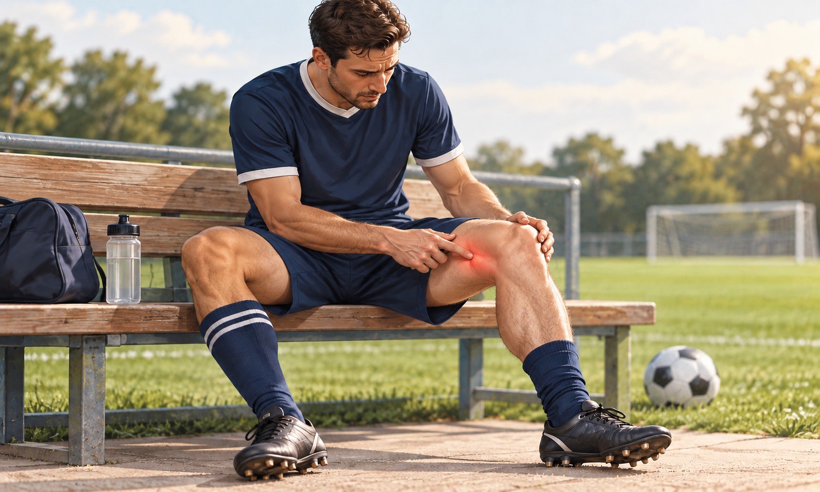
Physiotherapist assessing the inner knee of a patient with a suspected MCL injury in a New Malden clinic

MCL Injury

300+ Reviews 5.0

Open 7 Days

10+ Years' Experience

HCPC Physio On Team

Insurance Accepted

85% Return Rate

PHYSIOTHERAPY & THERAPEUTIC MASSAGE IN NEW MALDEN

Quick Summary

An MCL injury is a sprain or tear of the medial collateral ligament on the inner side of your knee, usually caused by a blow to the outside of the knee or a twisting force.

The great majority of isolated MCL injuries heal well without surgery, and our physiotherapists support your recovery with manual therapy and a targeted, criteria-based exercise programme.

Most people attend for a short course of sessions over several weeks. Recovery time depends on the grade of injury, ranging from around 1–2 weeks for a mild (Grade I) sprain to 6 weeks or more for a complete (Grade III) tear.

What Is an MCL Injury?

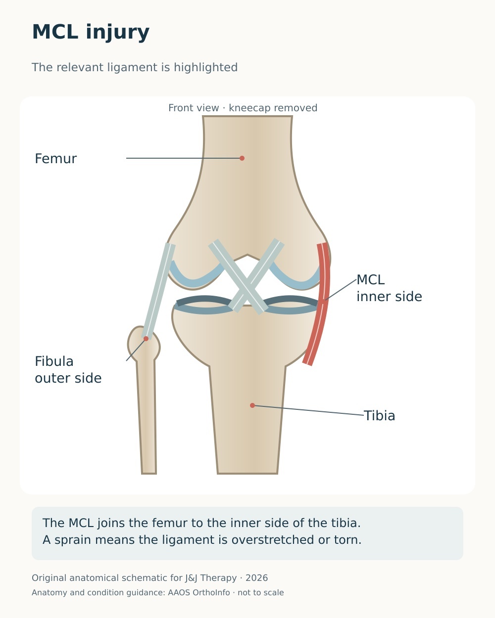
Anatomical diagram of the medial knee showing the superficial and deep MCL and posterior oblique ligament

The medial collateral ligament is the main structure resisting valgus (inward-bending) force on the knee, and it is the most commonly injured knee ligament. The medial side of the knee has a three-layer structure first described by Warren and Marshall (Warren & Marshall, Journal of Bone and Joint Surgery, 1979).


The key stabilisers are the superficial MCL — the largest component — the deep MCL (the meniscofemoral and meniscotibial fibres of the medial capsule), and the posterior oblique ligament. Detailed anatomical work has mapped the superficial MCL from its femoral origin just proximal and posterior to the medial epicondyle to two separate tibial insertions (LaPrade et al., Journal of Bone and Joint Surgery, 2007).


The MCL is injured in at least 42% of ligamentous knee injuries, with isolated MCL injuries accounting for around 29%. It is particularly common in contact and pivoting sports such as wrestling, judo, hockey and rugby (Roach et al., American Journal of Sports Medicine, 2014), and skiing is a leading civilian cause, with MCL injuries making up roughly 60% of knee injuries in skiers.


How injuries are graded

Grade I is a mild sprain with tenderness but no laxity. Grade II is a partial tear with some laxity but a firm endpoint. Grade III is a complete tear with marked laxity and a soft or absent endpoint. We assess using valgus stress at both full extension and 30 degrees of flexion, because laxity at 30 degrees alone suggests an isolated MCL injury, whereas laxity at both positions raises the possibility of additional cruciate ligament or posteromedial corner involvement.


Why the MCL usually heals well

Unlike the anterior cruciate ligament, the MCL sits largely outside the joint and has a rich blood supply, which supports a predictable healing sequence of bleeding, inflammation, repair and remodelling. Research suggests that controlled early movement produces better collagen alignment and greater tensile strength than prolonged immobilisation. A recent systematic review confirms that most isolated MCL injuries are managed without surgery, while noting that the supporting evidence is varied and of limited quality (Svantesson et al., BMJ Open Sport & Exercise Medicine, 2024).

Do You Experience These Symptoms?

✓  Pain and tenderness along the inner (medial) side of your knee

✓  Swelling over the inner knee, which is often mild because the ligament sits largely outside the joint

✓  A feeling that your knee may give way with side-to-side movement

✓  Stiffness and reduced range of movement

✓  Pain when the knee is pushed inwards, or when twisting and pivoting

✓  Bruising on the inner knee developing over the following days

✓  A pop or tearing sensation at the moment of injury in higher-grade tears

These symptoms can be treated.
The sooner you begin, the better your chances of a smoother recovery.

What Causes an MCL Injury?

•  Direct blow to the outer knee

A force to the outside of the knee, such as a lateral tackle in football or rugby, bends the knee inwards and overstretches the ligament.

This is the classic contact mechanism and often produces higher-grade injuries.


•  Twisting or cutting on a planted foot

Sudden changes of direction with the foot fixed create combined inward-bending and rotational stress across the inner knee.

This non-contact mechanism is common in pivoting sports and can injure the MCL on its own or alongside the anterior cruciate ligament.


•  Skiing falls

Skiing is a leading cause because the length of the ski increases the leverage placed through the inner knee during a fall.

Evidence shows MCL injuries account for around 60% of knee injuries sustained by skiers.


•  Combined higher-energy trauma

Greater forces can injure the MCL together with the anterior cruciate ligament, the medial meniscus and the posteromedial corner.

These combined patterns, and any suspected knee dislocation, need prompt medical assessment before rehabilitation begins.


•  Growth-related vulnerability in younger patients

In teenagers who are still growing, the growth plate at the lower end of the thigh bone is weaker than the ligament itself.

An injury that looks like an MCL sprain may therefore involve the growth plate, so we assess younger patients with particular care.

Not sure which condition applies to you?

How We Treat MCL Injury at J&J Therapy

Hands-On Manual Therapy

Our physiotherapists use manual therapy to ease pain, restore movement and support healing once your diagnosis and injury grade are clear. Techniques may include soft-tissue mobilisation, graded joint mobilisation and mobilisation with movement to recover full extension and flexion, alongside management of secondary muscular tightness. Because controlled loading rather than rest drives good ligament healing, hands-on treatment is paired with graded exercise from an early stage. We protect the inner knee from side-to-side stress in the early phase, and where indicated we advise a hinged knee brace, which may improve comfort and confidence while allowing early movement — though we are open with you that high-quality evidence for bracing is limited.

Shockwave Therapy*

For most MCL injuries, shockwave therapy is not used. Extracorporeal shockwave therapy is not recommended for a fresh (acute) ligament injury, where hands-on treatment and graded exercise are the priority. In a small number of cases, once the acute injury has fully settled but inner knee pain persists — for example with chronic ligament thickening, calcification at the ligament’s attachment (a Pellegrini-Stieda lesion) or associated soft-tissue pain — our physiotherapists may discuss the Swiss Storz Medical MASTERPULS (radial ESWT) as a second-line option after reassessment. The evidence here is limited to small studies and case reports, so we will always explain what shockwave can and cannot offer before recommending it.

Targeted Exercise Programme

Your targeted exercise programme is the core of MCL rehabilitation and follows a criteria-based progression rather than fixed dates. Early on we restore full extension, reactivate the quadriceps, help settle swelling and encourage weight-bearing as pain allows, because research suggests early controlled loading supports ligament healing. We then progress hip abductor, adductor and hamstring strengthening, followed by neuromuscular and balance training and, where appropriate, blood-flow-restriction training to build strength while protecting the healing ligament. Return to sport is guided by objective criteria — full pain-free range, restored strength and confident change of direction — with typical timeframes of around 1 to 2 weeks for Grade I, 3 to 4 weeks for Grade II and 6 weeks or more for Grade III.

When to Seek Urgent Medical Help

While most symptoms are not serious, urgent medical attention is required if you experience any of the following:

  • You cannot bear weight or a fracture is suspected — for example you cannot take four steps, or there is bony tenderness over the kneecap or the outer bump below the knee

  • Your knee is locked, cannot straighten, or feels grossly unstable in several directions, which may indicate a multi-ligament injury or knee dislocation

  • You have signs of a blood clot or infection — a hot, red, very swollen or feverish knee, or a painful swollen calf

  • A teenager who is still growing has inner knee pain after a valgus injury, as a growth-plate injury can mimic an MCL sprain and needs to be excluded

FAQs About MCL Injury

  • How long does an MCL injury take to heal?

It depends on the grade. A mild Grade I sprain often settles in around 1 to 2 weeks, a Grade II partial tear in roughly 3 to 4 weeks, and a complete Grade III tear in about 6 weeks or more. These are typical ranges — your physiotherapist will guide you using pain-free movement, strength and stability rather than the calendar, and the ligament continues to remodel quietly for many months afterwards.


  • Do I need a brace?

A hinged knee brace is often helpful for Grade II and Grade III injuries, protecting the ligament from side-to-side stress while still allowing you to move and strengthen the knee. It can improve comfort and confidence, but high-quality evidence for bracing is limited, so we treat it as one part of a wider plan rather than a substitute for exercise.


  • Can I walk on it, and when can I return to sport?

Most people can bear weight early, often with a brace or crutches at first, and early controlled movement actually helps healing. Return to sport is criteria-based: full pain-free range, restored strength and confident cutting and pivoting. As a rough guide this is around 1 to 2 weeks for Grade I, 3 to 4 weeks for Grade II and 6 weeks or longer for Grade III.


  • Will it heal without surgery?

Usually, yes. Most isolated MCL injuries, including many complete Grade III tears, recover well with non-operative treatment. Surgery is generally reserved for ongoing instability that has not settled with rehabilitation, certain tears at the shin-bone end of the ligament, and combined multi-ligament injuries.


  • Do you use shockwave therapy for an MCL injury?

Not for a fresh MCL injury. Shockwave therapy is not recommended during the acute phase, and hands-on treatment with graded exercise is the priority. In a small number of cases, once the injury has fully settled but inner knee pain persists — for example with calcification at the ligament’s attachment — we may discuss the Swiss Storz Medical MASTERPULS (radial ESWT) as a second-line option. The evidence is limited to small studies and case reports, and we will always be transparent about that before recommending it.

Not sure this condition matches your symtoms?
Find your condition and explore related pain areas.

Logo_Navy.png

Ready to start your recovery?
Book your Initial Assessment today.

This page is for general information only and does not replace professional medical advice.

Reviewed by Lavya Arigalayan, MSc, HCPC Registered Physiotherapist.

Last reviewed

2026년 8월 20일

bottom of page