Quick Summary
Knee cartilage injury describes damage to the smooth articular cartilage that lines the joint surfaces of your knee, causing pain, swelling and a catching sensation during movement. Because this tissue has no direct blood supply and very few cells, its capacity to repair itself is limited — which is why early, structured rehabilitation matters. Our physiotherapists typically recommend an initial course of 6–8 sessions, with your progress reviewed at every visit and your programme adjusted as your knee improves.
What Is a Knee Cartilage Injury?
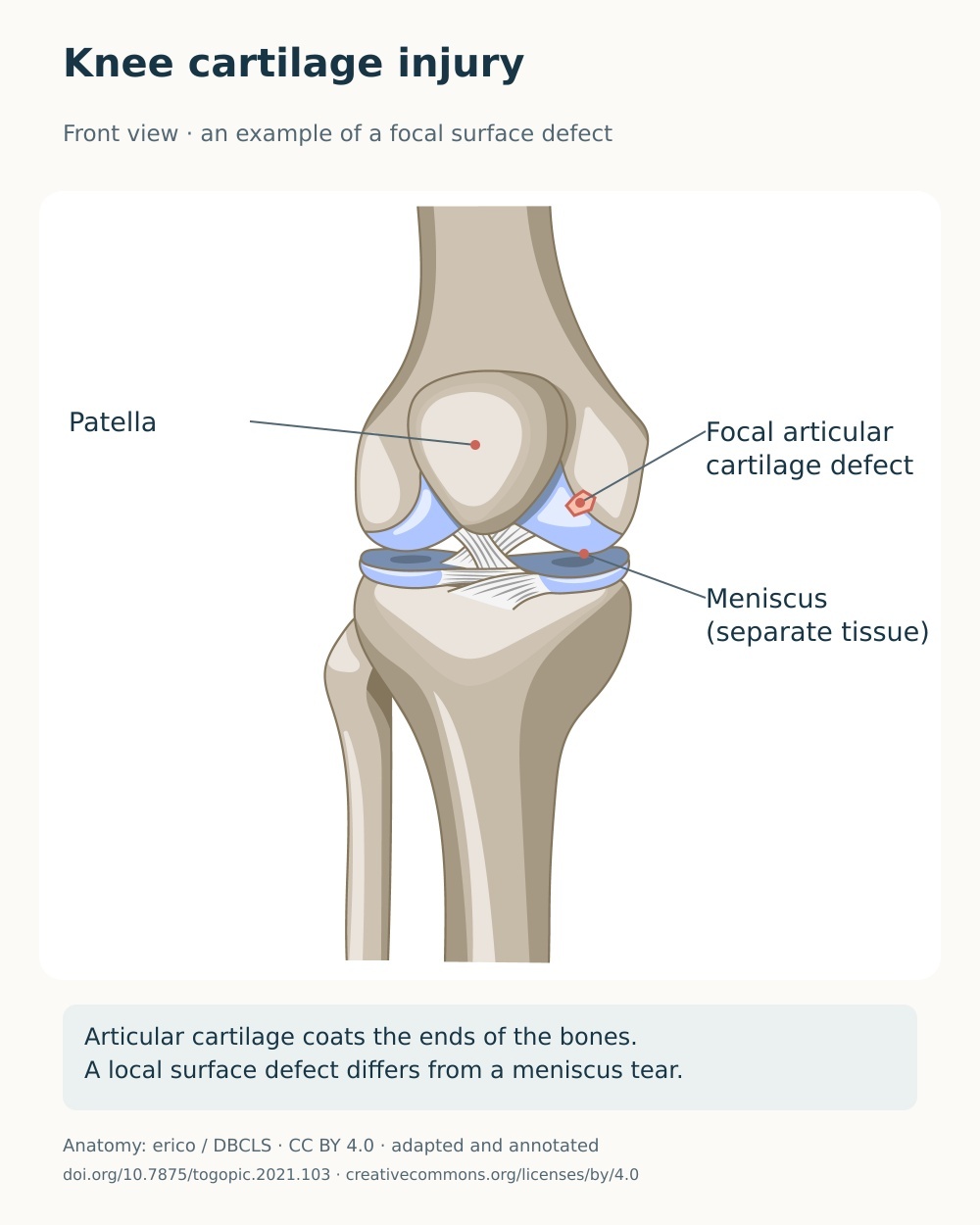
Articular cartilage is the glassy, low-friction tissue that caps the ends of the bones inside your knee — the femur, the tibia, and the back of your kneecap. It allows the joint to glide almost without friction and helps spread load evenly across the joint surfaces.
This tissue is avascular (it has no blood supply) and aneural (it has no nerve supply), and it contains relatively few cells. Research shows that because of these features it has a very limited ability to repair itself, and once damaged the tissue is often progressively lost — which can be a step towards osteoarthritis (Mistry et al., Clinics in Sports Medicine, 2018).
A chondral injury affects the cartilage alone, while an osteochondral injury involves the cartilage together with the bone beneath it. This distinction matters: because bone bleeds and cartilage does not, deeper osteochondral injuries trigger a repair response that fills the defect with fibrocartilage — a mechanically weaker tissue than the original hyaline cartilage — whereas damage confined to the cartilage surface may not heal at all.
How cartilage damage is graded
Clinicians grade the damage using the ICRS (International Cartilage Regeneration & Joint Preservation Society) or the closely related Outerbridge system, both running from Grade 0 to Grade IV:
• Grade I — the cartilage is intact but softened or swollen.
• Grade II — fraying and small fissures involving less than half the cartilage thickness.
• Grade III — deeper defects extending more than halfway through the cartilage, but not down to bone.
• Grade IV — full-thickness loss exposing the underlying bone.
The grade is only one part of the picture. A specialist also weighs the size of the defect, its location, your symptoms, and how your knee functions on examination.
Knee cartilage injury is related to — but distinct from — three other conditions we treat: meniscus tears, which affect the C-shaped fibrocartilage shock absorbers rather than the joint surface; chondromalacia patella, which is softening specifically behind the kneecap; and knee osteoarthritis, which is generalised, whole-joint wear. This page focuses on focal articular cartilage damage, meaning a discrete defect in an otherwise reasonably healthy joint.
Cartilage defects are found frequently at knee arthroscopy. In the largest series of its kind, Widuchowski et al. (The Knee, 2007) reviewed 25,124 knee arthroscopies and identified chondral lesions in 60% of patients, with Grade II changes the most common finding. Focal cartilage injury is strongly associated with anterior cruciate ligament (ACL) rupture and meniscal tears, and is more common in athletes and in younger, active people following a twisting or impact injury.
Do You Experience These Symptoms?
✓ Deep, poorly localised knee pain that is worse with weight-bearing, squatting, stairs or impact
✓ Swelling that comes and goes, often appearing a few hours after activity
✓ Catching, clicking or a sensation of something being caught inside the joint
✓ Locking, or the knee giving way, particularly if a fragment has become loose
✓ Stiffness and reduced range of movement after rest or after activity
✓ Tenderness over the joint line or over the affected joint surface
✓ A grinding sensation (crepitus) as you bend and straighten the knee
These symptoms can be treated.
The sooner you begin, the better your chances of a smoother recovery.
What Causes Knee Cartilage Injury?
• Acute traumatic injury
A direct blow, a fall onto the knee, or a twisting and pivoting injury can shear off or bruise a section of cartilage.
These injuries frequently occur alongside ligament or meniscal damage, particularly with ACL ruptures and kneecap dislocations.
• Sports and repetitive high-impact loading
Running, jumping, football, skiing and other pivoting sports place large, repeated forces across the joint surfaces.
Over time this can produce focal wear or a fatigue-type lesion, and cartilage defects are notably more common in athletes than in the general population.
• Associated knee injuries and instability
A deficient ACL or a torn meniscus alters how load is transmitted through the knee, exposing the cartilage to abnormal shear and contact forces.
This is why an unstable knee carries a higher risk of developing progressive cartilage damage over time.
• Osteochondritis dissecans
In some people, usually adolescents and young adults, a segment of bone just beneath the cartilage loses its blood supply and can partially or fully separate along with its overlying cartilage.
If the fragment detaches it becomes a loose body within the joint, causing locking and other mechanical symptoms.
• Malalignment and uneven load distribution
Bow-legged or knock-kneed alignment, or a kneecap that tracks poorly, concentrates force on one part of the joint.
This uneven loading can accelerate focal cartilage breakdown in the overloaded area.
How We Treat Knee Cartilage Injury at J&J Therapy
Hands-On Manual Therapy
Patellofemoral and tibiofemoral joint mobilisation with targeted soft-tissue work to the quadriceps, hamstrings and calf, alongside treatment of contributing areas at your hip and ankle.
Research suggests that combining manual therapy with exercise improves pain and function more than exercise alone (Deyle et al., Annals of Internal Medicine, 2000).
Hands-on treatment helps settle pain and stiffness so you can load the knee more comfortably and get the most from your strengthening work.
Shockwave Therapy*
Swiss Storz Medical MASTERPULS
may be considered as a second-line adjunct where your assessment identifies associated peri-articular soft-tissue or bone-stress symptoms — it is not applied to the cartilage defect itself, and evidence in this specific condition remains limited.
Non-invasive, and only used when your physiotherapist judges it appropriate alongside your manual therapy and exercise programme.
Not suitable for younger patients with open growth plates in the treatment area.
Targeted Exercise Programme
A progressive, individualised programme built around load management, quadriceps and hip strengthening, and neuromuscular control — the strongest evidence-based element of your care.
We combine closed-chain and carefully graded open-chain work, and where heavier loading is not yet comfortable we may use low-load blood flow restriction training (Hughes et al., British Journal of Sports Medicine, 2017).
Return to sport is guided by objective strength and hop-test criteria rather than time alone.
When to Seek Urgent Medical Help
While most symptoms are not serious, urgent medical attention is required if you experience any of the following:
A locked knee — a sudden inability to fully straighten your knee, which may indicate a loose fragment blocking the joint
A significant injury with an audible pop followed by rapid, marked swelling within the first few hours
Complete inability to bear weight, or an obviously deformed knee, after trauma
A hot, red, very swollen knee with fever or feeling generally unwell — this needs same-day medical care
FAQs About Knee Cartilage Injury
Can knee cartilage heal on its own?
Articular cartilage has a very limited ability to repair itself because it has no blood supply and contains few cells. Research shows that damage confined to the cartilage surface often does not heal, while deeper injuries that reach the bone may fill with a weaker, fibrocartilage-type tissue. This is why we focus on protecting the joint, building strength and managing load rather than expecting the cartilage to regrow.
Do I need surgery for a cartilage injury?
Not always. Many focal cartilage injuries are managed successfully without surgery, and NICE guidance recommends a trial of physiotherapy, load modification and weight management first. Surgery is generally considered only when symptoms persist despite well-structured conservative care, or when a lesion is large, unstable, or causing mechanical locking. If we identify those features, we will refer you for orthopaedic assessment.
How long will my rehabilitation take?
This varies with the size and location of the injury and with your goals. Many people notice improvements in pain and swelling within a few weeks, but rebuilding strength and returning confidently to sport is usually a three to six month process. We review your progress at every session and adjust your plan accordingly.
Will exercise make the damage worse?
No — appropriately dosed exercise is protective rather than harmful. The key is correct loading: too much too soon can flare your symptoms, but well-graded strengthening improves how your joint tolerates load and reduces pain. Our physiotherapists tailor the intensity to your symptoms and progress it safely.
Can shockwave therapy repair my cartilage?
No, and we would never present it that way. There is currently no robust trial evidence that shockwave therapy repairs knee cartilage — most of the available evidence relates to knee osteoarthritis and to bone and soft-tissue problems rather than to focal cartilage defects, and our radial device cannot target the deep cartilage surface directly. We only consider it after a full assessment, as a second-line adjunct for associated peri-articular or bone-stress symptoms alongside your exercise programme, and not for patients with open growth plates.
This page is for general information only and does not replace professional medical advice.
Reviewed by Lavya Arigalayan, MSc, HCPC Registered Physiotherapist.
Last reviewed:
2026년 8월 20일