Quick Summary
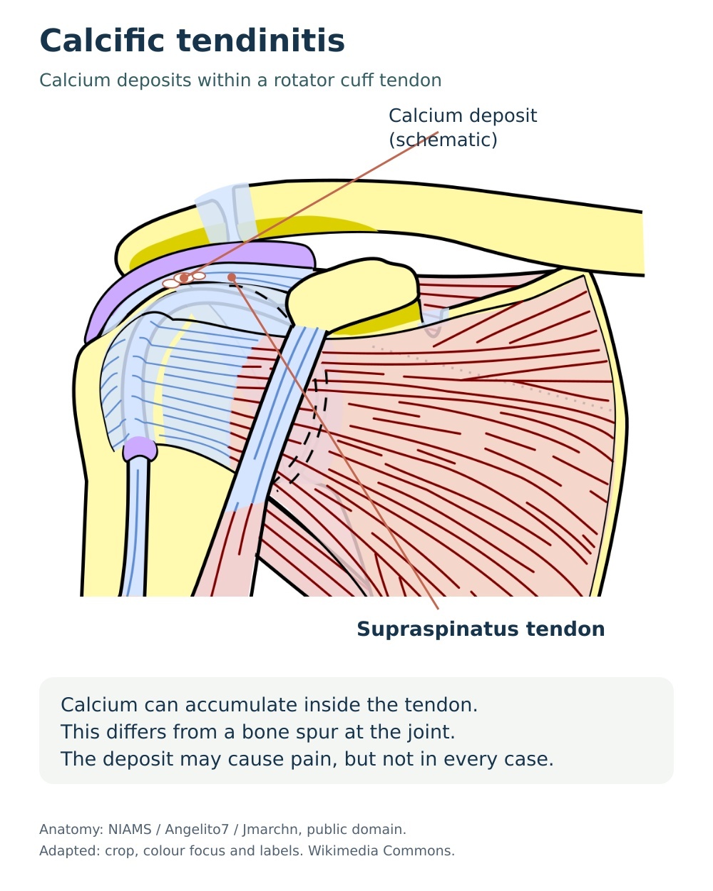
Calcific tendinitis is a common and often intensely painful shoulder condition in which calcium deposits form within one of your rotator cuff tendons — most often the supraspinatus at the top of the shoulder.
It is usually self-limiting, and research suggests that most people improve with non-surgical care. Shockwave therapy is one of the most strongly evidence-supported options, and many patients improve over a typical course of 3–6 sessions alongside targeted rehabilitation.
At J&J Therapy in New Malden, our HCPC-registered physiotherapists combine manual therapy, Swiss Storz Medical shockwave therapy and a progressive exercise programme to help reduce your pain, restore your movement and support your body's natural resorption of the deposit.
What Is Calcific Tendinitis?
Calcific tendinitis — also called calcific tendinopathy of the rotator cuff — occurs when calcium hydroxyapatite crystals are deposited within one or more of the tendons that stabilise your shoulder. The supraspinatus tendon is involved in the large majority of cases, followed by the infraspinatus and, less commonly, the subscapularis (Louwerens et al., Journal of Shoulder and Elbow Surgery, 2015).
How common is it?
Calcific deposits are far more common than many people realise. In a clinical and radiological analysis of over 1,200 adults, deposits were found in around 7.8% of people with no shoulder symptoms and in 42.5% of those presenting with subacromial shoulder pain (Louwerens et al., 2015). It most often affects adults between the ages of 30 and 60, and is reported more frequently in women than in men.
Why the deposits form
The most widely accepted explanation is that this is an active, cell-mediated process rather than simple wear and tear. A small area of tendon undergoes a change in cell type in a low-oxygen environment, and these altered cells lay down calcium (Uhthoff and Sarkar, Clinical Orthopaedics and Related Research, 1976; Uhthoff and Loehr, Journal of the American Academy of Orthopaedic Surgeons, 1997).
The three stages
• Precalcific stage: the tendon tissue changes character, usually without symptoms.
• Calcific stage: calcium is laid down (formative phase), sits quietly and often painlessly (resting phase), then begins to break down as the body sends in blood vessels and clean-up cells (resorptive phase). The resorptive phase is when pain is typically at its most severe.
• Postcalcific stage: the deposit clears and the tendon remodels and heals.
What this means for you
Because calcific tendinitis has a strong natural tendency towards resorption, many people settle with conservative care — but the timescale is unpredictable and can range from weeks to well over a year. Two typical patterns are recognised: a chronic, catching ache with overhead activity, and a sudden, severe pain of abrupt onset during the resorptive phase (Speed and Hazleman, New England Journal of Medicine, 1999). The aim of treatment is to control your pain, restore your function and, where appropriate, support the breakdown of the deposit.
Do You Experience These Symptoms?
✓ Shoulder pain that may be a chronic dull ache or come on suddenly and severely, often without any injury
✓ Pain felt mainly at the front and outer side of your shoulder, sometimes spreading into your upper arm
✓ Night pain that disturbs your sleep, and difficulty lying on the affected side
✓ Pain or catching when you lift your arm overhead, reach up or reach behind your back
✓ Stiffness and reduced range of movement, particularly when lifting the arm out to the side
✓ A sense of weakness or reluctance to use the arm because of pain
✓ During an acute flare, intense pain that makes almost any shoulder movement difficult
These symptoms can be treated.
The sooner you begin, the better your chances of a smoother recovery.
What Causes Calcific Tendinitis?
• Tendon cell changes and reduced blood supply
The leading theory is that a poorly supplied area of tendon changes cell type in a low-oxygen environment, and these altered cells begin depositing calcium.
This is regarded as an active biological process rather than straightforward wear and tear, which is why it can occur in otherwise healthy shoulders.
• Age and sex
Calcific tendinitis most commonly develops between the ages of 30 and 60 and becomes less typical outside this range.
It is consistently reported to be more common in women, who make up the majority of symptomatic cases.
• Metabolic and thyroid conditions
Diabetes and thyroid disorders are associated with a higher likelihood of developing calcific tendinitis.
Research suggests that people with these conditions may develop symptoms at a younger age and experience a longer course (Harvie et al., Journal of Shoulder and Elbow Surgery, 2007).
• Individual and familial predisposition
A familial tendency has been described, suggesting that some people are simply more predisposed to forming calcific deposits.
This helps explain why the condition often appears with no obvious mechanical trigger at all.
• Repetitive overhead loading
Repeated overhead work or sport places cumulative demand on the rotator cuff and may contribute to the tendon changes that precede calcification.
Mechanical load is only part of the picture, however, as many people develop the condition without any history of overuse.
How We Treat Calcific Tendinitis at J&J Therapy
Hands-On Manual Therapy
Our physiotherapists use manual therapy to help settle your pain and restore movement while the deposit resolves. Depending on how irritable your shoulder is, we may use gentle joint mobilisations, soft-tissue techniques for the surrounding muscles, and graded movement work to improve how your shoulder blade and shoulder joint work together. During an acute flare, treatment is deliberately gentle and guided by your pain, with the focus on settling symptoms and protecting your sleep; as your pain eases we progress hands-on work to help you regain comfortable overhead reach. Manual therapy is delivered as part of a combined programme rather than on its own, in line with guidance that shockwave therapy should be provided alongside physiotherapy (NICE, IPG742, 2022).
Shockwave Therapy*
Swiss Storz Medical MASTERPULS radial shockwave delivers targeted acoustic pulses to the affected rotator cuff tendon, helping to fragment calcium deposits and stimulate their natural resorption while reducing local pain sensitivity. Research shows shockwave therapy can improve shoulder pain and function and promote breakdown of the deposit. Non-invasive, evidence-based. Typically 3–6 sessions.
Targeted Exercise Programme
Once your pain allows, we guide you through a progressive exercise programme built around your stage and your goals. Early on we focus on gentle range-of-movement and postural work to keep your shoulder mobile without provoking the deposit. As symptoms settle, we add graded rotator cuff and shoulder blade strengthening to restore stability, control and comfortable overhead function. Your home programme is central to progress — UK shoulder care guidance supports a structured course of physiotherapy and exercise as first-line management, and consistent practice between sessions is what tends to drive lasting improvement. We adjust the plan if you move into an acute flare.
When to Seek Urgent Medical Help
While most symptoms are not serious, urgent medical attention is required if you experience any of the following:
A red, hot or swollen shoulder with fever or feeling generally unwell — this may indicate joint infection and needs same-day emergency assessment
Sudden loss of movement or marked weakness after an injury, such as being unable to lift your arm — this may indicate a tendon tear or fracture and needs urgent review
A new lump or swelling around the shoulder, or new unexplained shoulder pain if you have a history of cancer — arrange urgent medical review
Severe pain that is not controlled by your usual pain relief, or shoulder pain with chest pain or breathlessness — seek prompt medical advice
FAQs About Calcific Tendinitis
Will calcific tendinitis go away on its own?
Often, yes. Calcific tendinitis is usually self-limiting and the body tends to resorb the calcium deposit over time. The timescale is unpredictable and the pain can be severe, so treatment aims to control your symptoms, restore your movement and support that natural process rather than simply waiting it out.
How is calcific tendinitis diagnosed?
It is normally diagnosed from your history and a physical examination, supported by imaging. An X-ray or ultrasound scan will usually show the calcium deposit and can help indicate which phase it is in. Our physiotherapists can assess your shoulder and, where appropriate, recommend onward referral for imaging.
Does shockwave therapy work for calcific tendinitis?
Evidence shows that shockwave therapy can help. In a double-blind randomised controlled trial, shockwave therapy improved shoulder function substantially more than sham treatment and was associated with disappearance of the deposit in a far higher proportion of patients (Gerdesmeyer et al., JAMA, 2003), and a systematic review and meta-analysis confirmed improvements in pain, function and calcium resorption at six months (Ioppolo et al., Archives of Physical Medicine and Rehabilitation, 2013). Most people are treated over a course of around 3–6 sessions, used alongside manual therapy and exercise. We will always be open with you about what the evidence does and does not show — NICE regards the overall efficacy evidence as still developing and advises that shockwave be delivered together with physiotherapy (NICE, IPG742, 2022).
What if physiotherapy and shockwave therapy do not settle my symptoms?
For persistent cases, your GP or consultant may discuss an ultrasound-guided corticosteroid injection for short-term pain relief, ultrasound-guided needling and lavage to break up and wash out the deposit, or, rarely, keyhole surgery to remove it. These are generally considered when symptoms do not settle with conservative care, and we are happy to explain the options and liaise with your doctor.
How long will recovery take?
This varies considerably because the condition naturally fluctuates. Some people settle within weeks to a few months, while longer-standing cases can take more time. We will set realistic expectations for your particular presentation and adjust your programme as you progress.
This page is for general information only and does not replace professional medical advice.
Reviewed by Lavya Arigalayan, MSc, HCPC Registered Physiotherapist.
Last reviewed:
2026년 8월 20일